Overview
The navicular bone of the foot is one of the small bones on the mid-foot. The bone is located at the instep, the arch at the middle of the foot. One of the larger tendons of the foot, called the posterior tibial tendon, attaches to the navicular before continuing under the foot and into the forefoot. This tendon is a tough band of tissue that helps hold up the arch of the foot. If there is an accessory navicular, it is located in the instep where the posterior tibial tendon attaches to the real navicular bone.
Causes
An injury to the fibrous tissue connecting the two bones can cause something similar to a fracture. The injury allows movement to occur between the navicular and the accessory bone and is thought to be the cause of pain. The fibrous tissue is prone to poor healing and may continue to cause pain. Because the posterior tibial tendon attaches to the accessory navicular, it constantly pulls on the bone, creating even more motion between the fragments with each step.
Symptoms
The catalyst for symptoms might be some sort of injury (such as a broken or sprained ankle), excessive activity, or irritation from where shoes are rubbing on the bony prominence the accessory navicular What makes you grow taller during puberty?. These can irritate the bone, or make the tendon it?s embedded in (the posterior tibial tendon, remember?) inflamed and sore. Because the tendon is compromised in its ability to support the arch, accessory navicular syndrome almost always leads to flat feet, which is one very obvious symptom.
Diagnosis
Diagnosis starts by speaking with the patient about symptoms. The physician will look at the foot and examine it for signs of an accessory navicular. By putting pressure on the area, the doctor may determine its presence simply by the presence of pain. The muscle, joint, and the overall structure of the foot may be considered, as well as the way in which the patient walks. If a diagnosis of accessory navicular syndrome is made, an X-ray or MRI may be ordered to confirm diagnosis.
Non Surgical Treatment
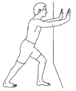
The initial treatment approach for accessory navicular is non-operative. An orthotic may be recommended or the patient may undergo a brief period of casting to rest the foot. For chronic pain, however, the orthopedic surgeon removes the extra bone, a relatively simple surgery with a brief rehabilitation period and a very good success rate.

Surgical Treatment
The original procedure advocated by Kidner involved shelling out of the accessory navicular bone from within the insertional area of the posterior tibial tendon and rerouting this tendon under the navicular bone in hopes of restoring a normal pull of this tendon. When treating younger children, history has shown us that simply shelling out of the accessory navicular bone from within the tendon and remodeling the tuberosity of the navicular bone can give you satisfactory results.
In general, you want to reserve advancement of the posterior tibial tendon for adults or those who have a more significant flatfoot deformity. You may also use this approach after determining that quality custom orthotics are only resulting in a slight decrease of symptoms.
Th1s1sanart1cl3s1te
The navicular bone of the foot is one of the small bones on the mid-foot. The bone is located at the instep, the arch at the middle of the foot. One of the larger tendons of the foot, called the posterior tibial tendon, attaches to the navicular before continuing under the foot and into the forefoot. This tendon is a tough band of tissue that helps hold up the arch of the foot. If there is an accessory navicular, it is located in the instep where the posterior tibial tendon attaches to the real navicular bone.
Causes
An injury to the fibrous tissue connecting the two bones can cause something similar to a fracture. The injury allows movement to occur between the navicular and the accessory bone and is thought to be the cause of pain. The fibrous tissue is prone to poor healing and may continue to cause pain. Because the posterior tibial tendon attaches to the accessory navicular, it constantly pulls on the bone, creating even more motion between the fragments with each step.
Symptoms
The catalyst for symptoms might be some sort of injury (such as a broken or sprained ankle), excessive activity, or irritation from where shoes are rubbing on the bony prominence the accessory navicular What makes you grow taller during puberty?. These can irritate the bone, or make the tendon it?s embedded in (the posterior tibial tendon, remember?) inflamed and sore. Because the tendon is compromised in its ability to support the arch, accessory navicular syndrome almost always leads to flat feet, which is one very obvious symptom.
Diagnosis
Diagnosis starts by speaking with the patient about symptoms. The physician will look at the foot and examine it for signs of an accessory navicular. By putting pressure on the area, the doctor may determine its presence simply by the presence of pain. The muscle, joint, and the overall structure of the foot may be considered, as well as the way in which the patient walks. If a diagnosis of accessory navicular syndrome is made, an X-ray or MRI may be ordered to confirm diagnosis.
Non Surgical Treatment
The initial treatment approach for accessory navicular is non-operative. An orthotic may be recommended or the patient may undergo a brief period of casting to rest the foot. For chronic pain, however, the orthopedic surgeon removes the extra bone, a relatively simple surgery with a brief rehabilitation period and a very good success rate.
Surgical Treatment
The original procedure advocated by Kidner involved shelling out of the accessory navicular bone from within the insertional area of the posterior tibial tendon and rerouting this tendon under the navicular bone in hopes of restoring a normal pull of this tendon. When treating younger children, history has shown us that simply shelling out of the accessory navicular bone from within the tendon and remodeling the tuberosity of the navicular bone can give you satisfactory results.
In general, you want to reserve advancement of the posterior tibial tendon for adults or those who have a more significant flatfoot deformity. You may also use this approach after determining that quality custom orthotics are only resulting in a slight decrease of symptoms.
Th1s1sanart1cl3s1te